
Down syndrome (Trisomy 21), Edward syndrome (Trisomy 18), Patau syndrome (Trisomy 13) and neural tube defects (ONTD) are commonly considered fatal or dangerous genetic defects for the fetus. These diseases are classified as chromosomal and genetic abnormalities.
Amniocentesis and CVS are not only very expensive in terms of the cost of tests, but because of the invasive method of obtaining samples (amniotic fluid as well as placental villi) can cause miscarriage, so the use of these two methods except in very cases Not recommended.
In comparison, screening tests are non-invasive and cost-effective. Also, due to the use of blood samples from pregnant mothers, it is possible to repeat these tests in the same week or in subsequent weeks (from week 11-17 of pregnancy).
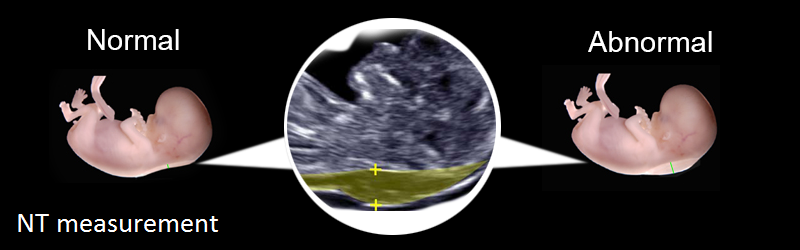
At present, in addition to maternal age, race, gestational age, maternal weight, diabetes, smoking, history of abortion, use of IVF and IUI methods for pregnancy, number of fetuses, sonographic parameters including BPD, CRL, NT, NB and detailed report Fetal age is also measured using laboratory parameters including serum levels of AFP, Unconjugated E3, Inhibin-A, BHCG, PAPP-A and Free BHCG.
Obviously, all the above parameters should lead to a risk report using a standard and international software that has a suitable statistical algorithm.
Today, in addition to holding the risk of the mentioned diseases, the risk of preeclampsia (pregnancy poisoning) as well as the risk of the rare Smith - Lemli - Opitz. Syndrome (SLOS) can also be measured and reported.
Ductus Venosus pulsatility index (DVPI)
What is DVPI?
DVPI is a quantitative indicator that can be easily measured by ultrasound.
When to do DVPI?
Add DVPI to Double marker, Combined test, Integrated test
At 10-13 weeks of gestation, the effectiveness of the prenatal screening test for Down syndrome is significantly increased and the number of false positives is reduced by up to 50% at a certain diagnosis.
Impact of DVPI on screening?
In a fetus with Down syndrome, DV blood flow may be abnormal. In this case, a slight increase in the index is evident in ultrasound. Recent studies show that DV blood flow at the end of diastole is reversed in 64% of pregnancies with Down syndrome and 4% of normal pregnancies.
(Normally, DV blood flows from the placenta to the fetus, while in Down syndrome, it flows from the fetus to the placenta).
According to Median, DVPI is 1.71 in pregnancies with Down syndrome, while it is 1.1 in normal pregnancies.
What we need to know about screening:
- All pregnant women, regardless of age, should be screened for the most common anoploids.
- Diagnostic tests should be available to all women who present before 18 weeks of gestation. Pregnant women should be informed of the differences between screening and diagnostic methods.
- In accordance with the latest international standards and the country's health reference, to achieve the most accurate results, the most appropriate time to perform screening is the first trimester of pregnancy, 11 to 13w + 6day and the second trimester, 14 to 17 weeks of pregnancy.
- Ideally, for those who visit in the first trimester, it is best to use screening methods that include the first and second trimesters (sequential, integrated).
- Methods that use a combination of first and second-trimester screening, such as Sequential and Integrated, are preferred over other methods because of their higher detection rates.
- Women who have screening tests in the first trimester should be tested at least for NTD (AFP) screening in the second trimester.
- Smith Lemli Opitz (SLOS) Syndrome is an autosomal recessive disorder associated with mental retardation with a minimum prevalence of 1 in 60,000. In this syndrome, the amount of cholesterol is abnormal and the concentration of cholesterol and its precursors in the blood is low. This syndrome can be diagnosed before delivery by high levels of amniotic fluid 7 - dehydrocholesterol. In pregnancies where the fetus has SLOS, the serum level of maternal AFP, such as UE3 and hCG, is low. The risk of this abnormality can be reported by screening test.